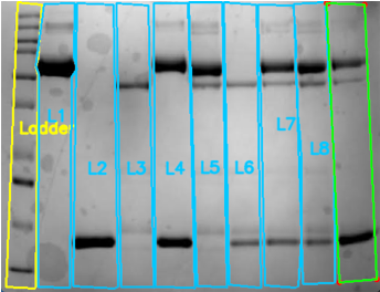
Research

Multiple Projects with one unifying question:
How can we make our science and our medicine better?

PROJECT 01

The development of the Venetian Circulation for biophysical simulation of hemodynamics.

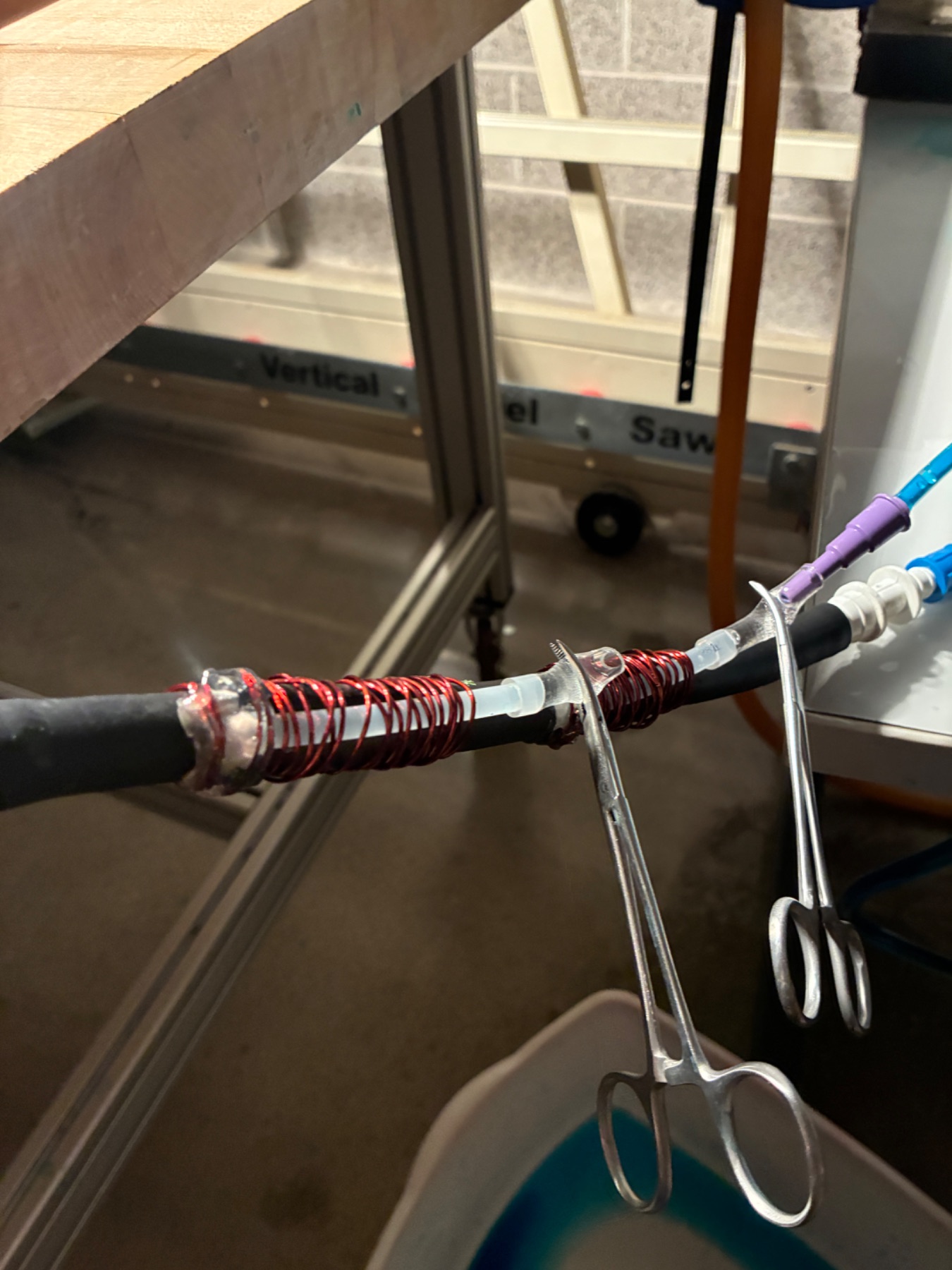
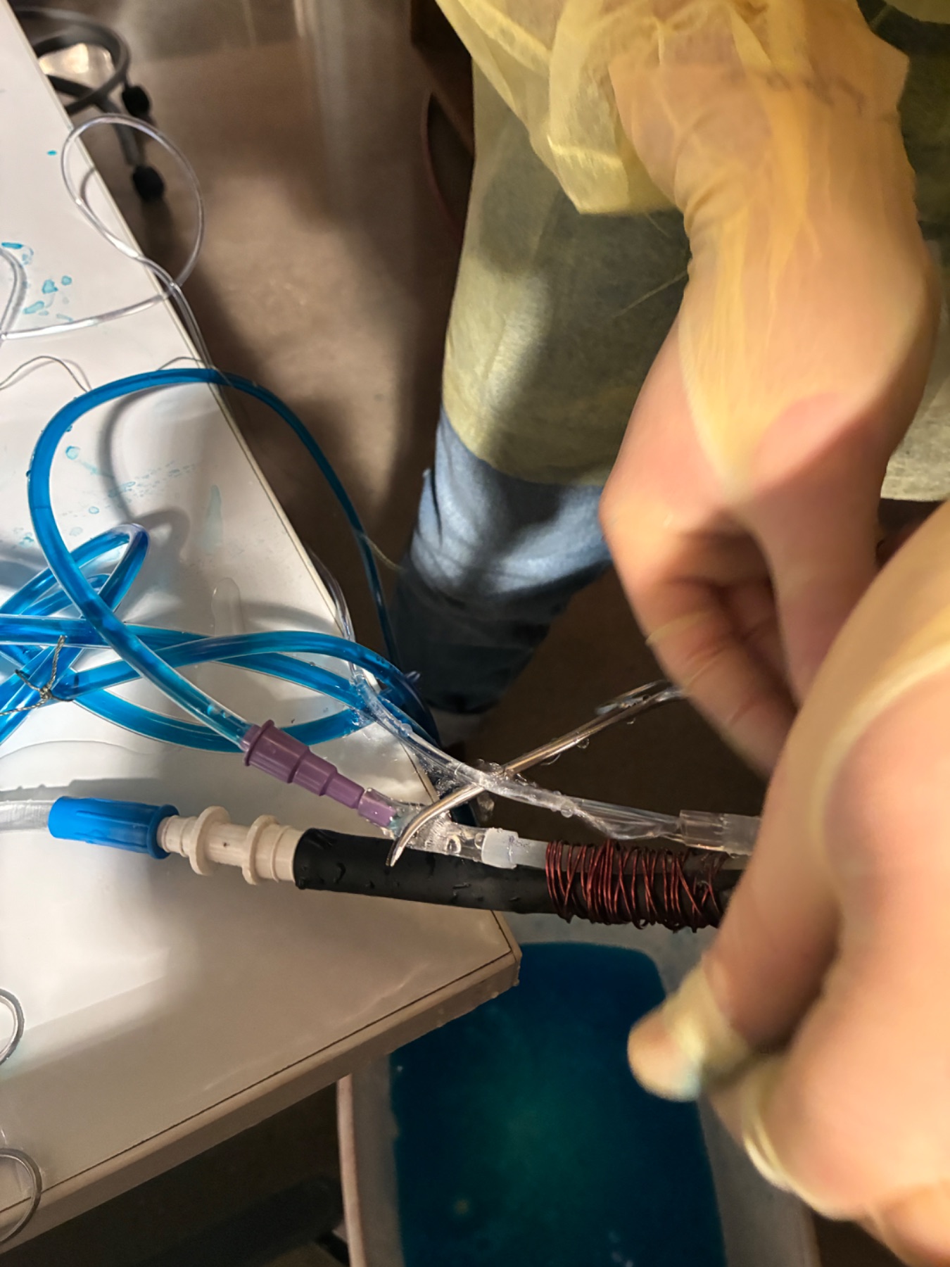
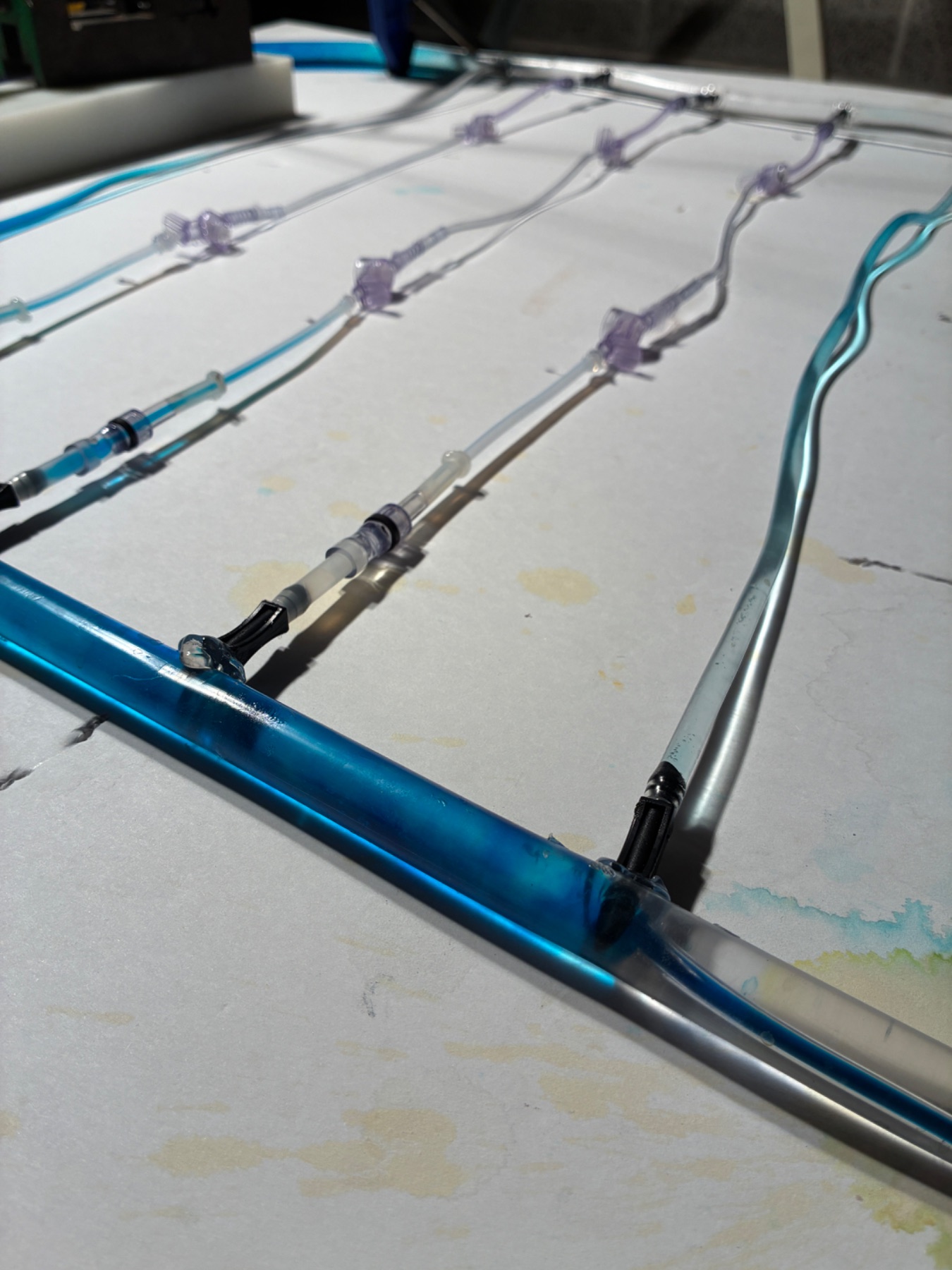
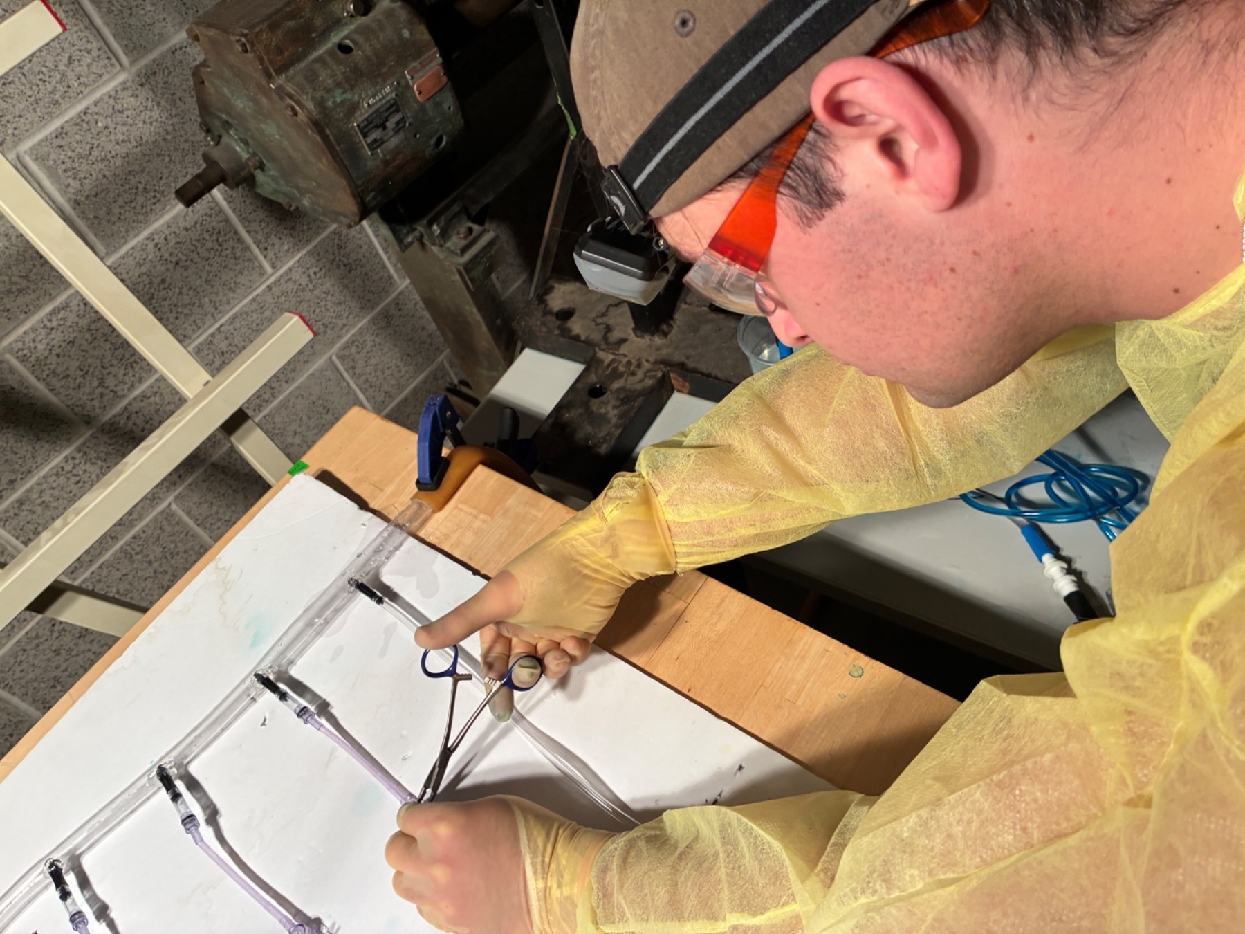
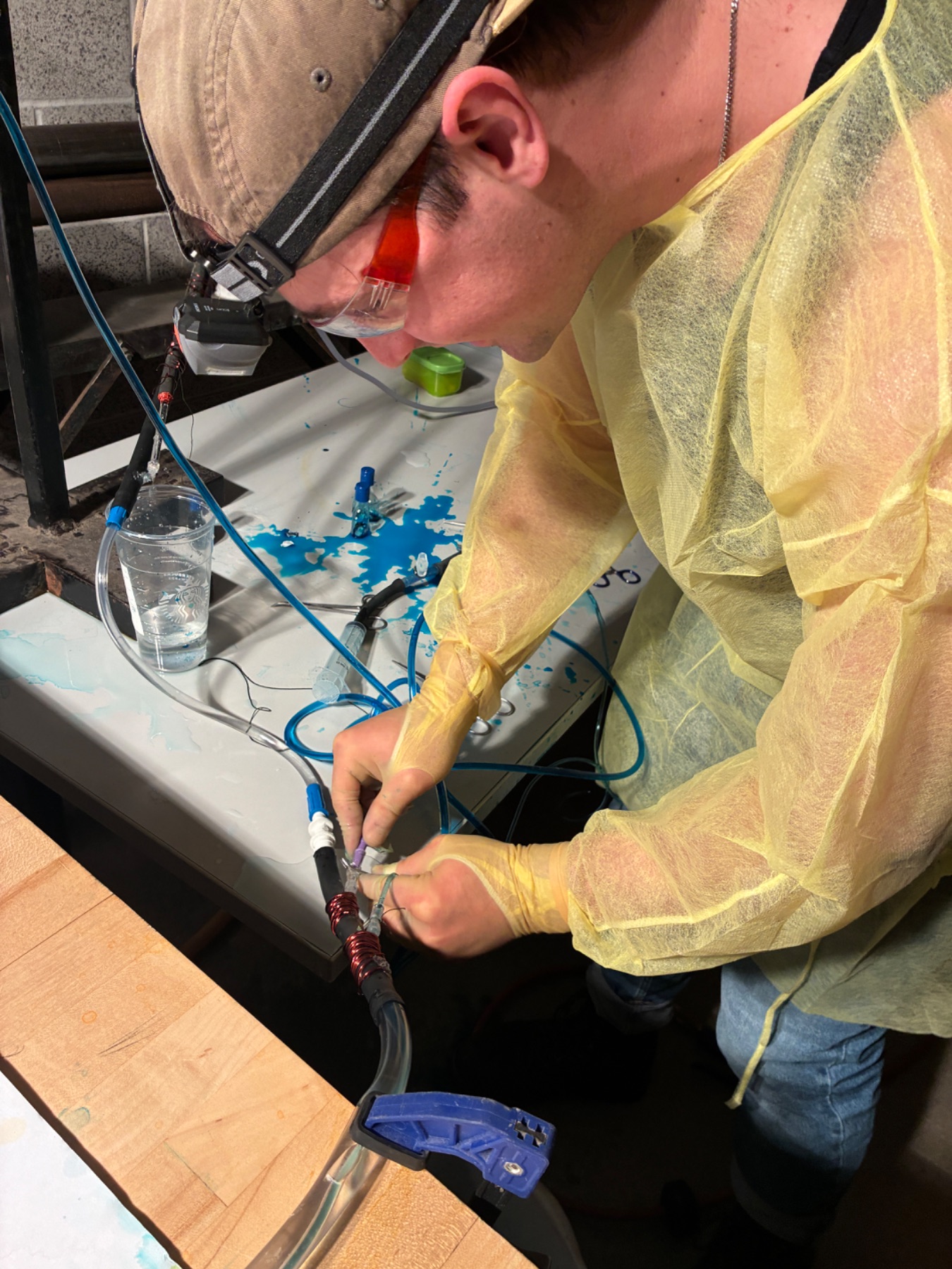
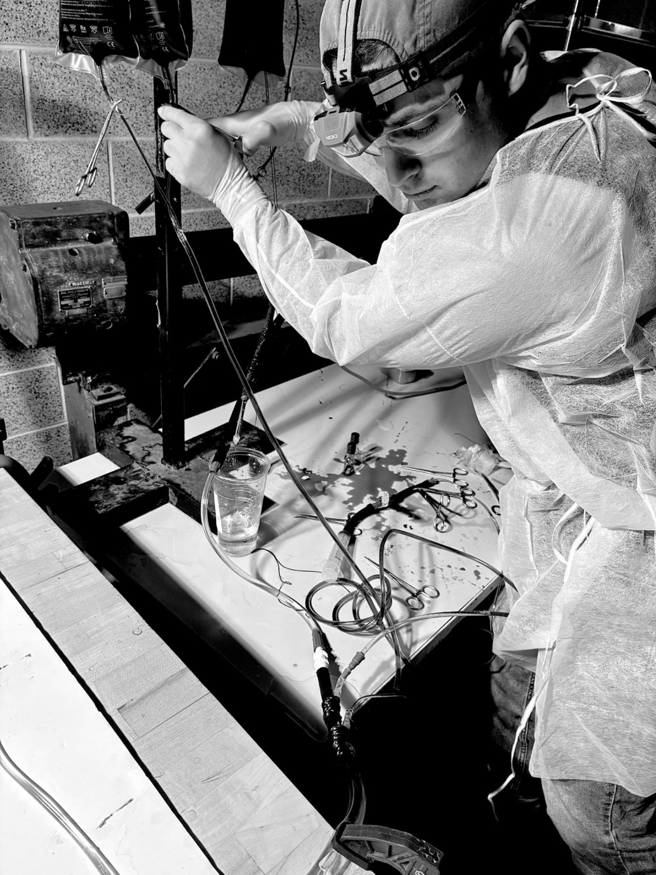
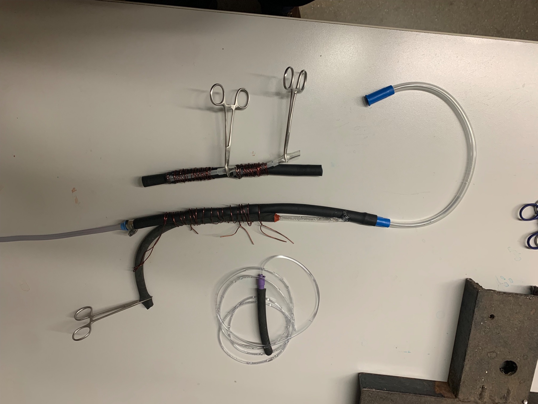
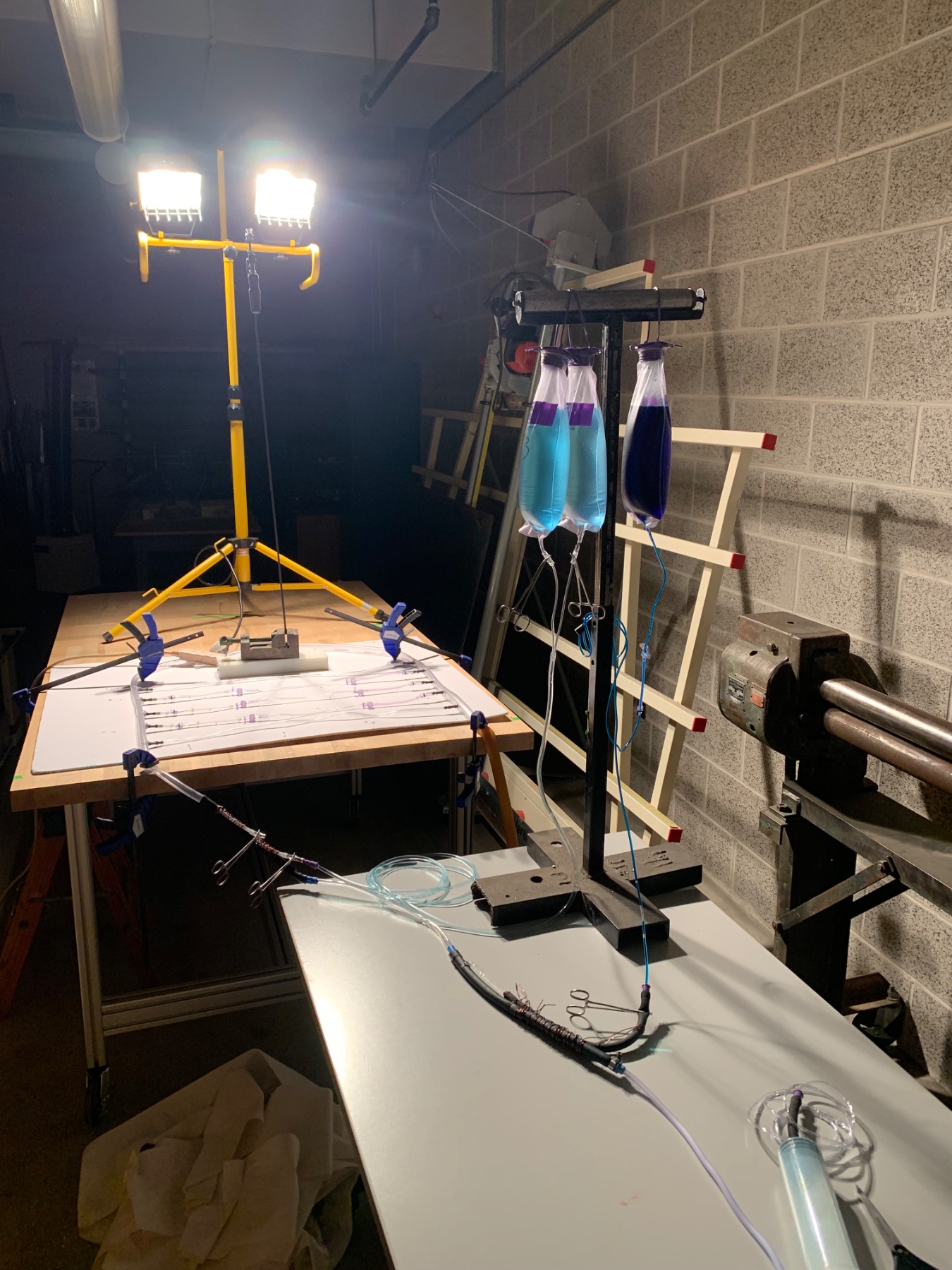
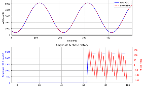
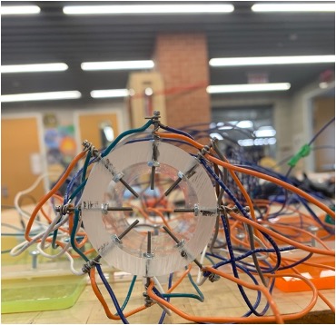
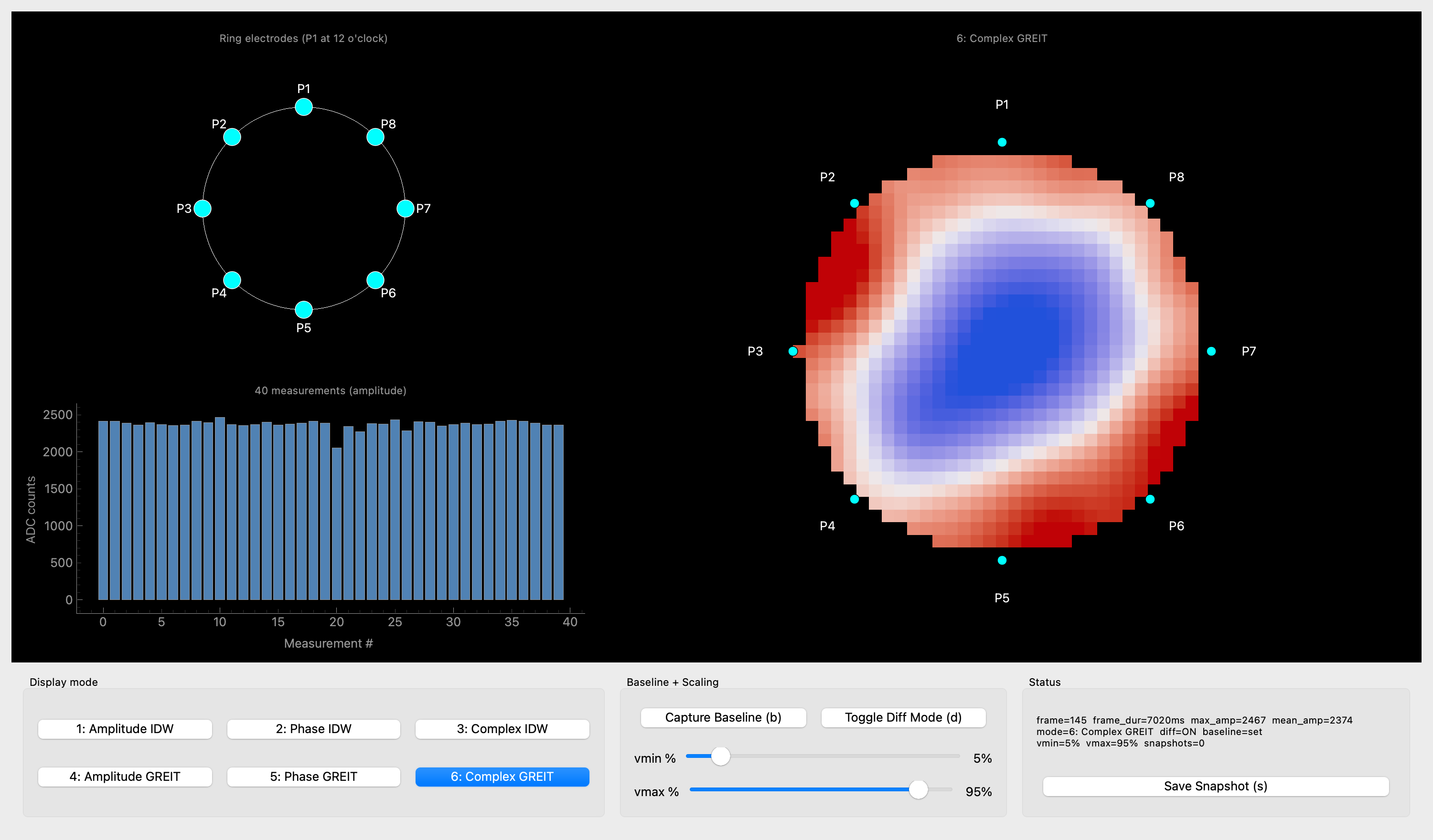
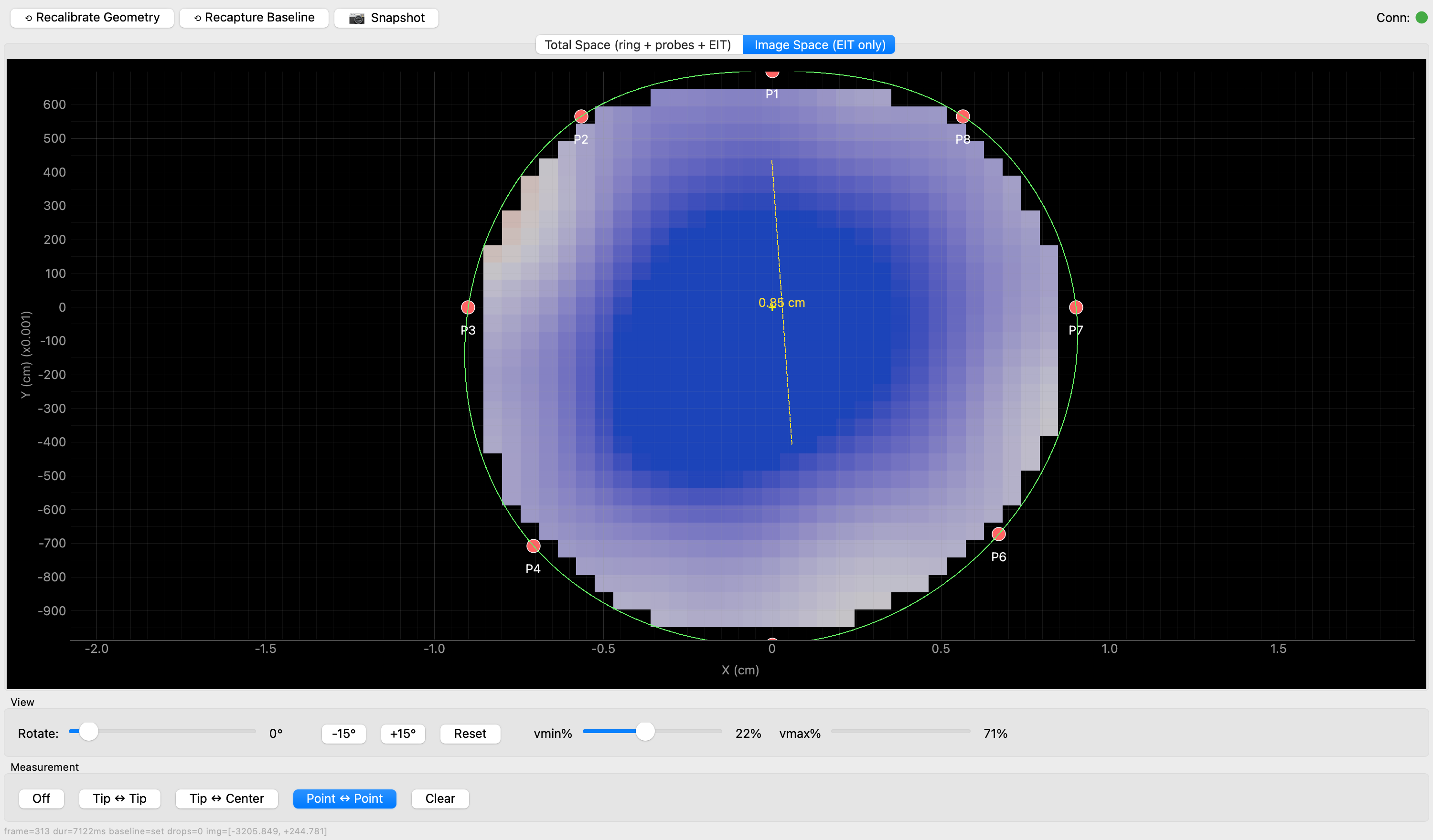
The Venetian Circulation is a biophysical simulation of the circulatory system capable of fluid-dynamic measurement through optical angiography. The device is currently being used to simulate and study decompensated shock, probing for potential treatment strategies. In addition, its angiography capabilities are being adapted to simulate fluoroscopy — an adaptation that could become the foundation for further innovation in PCI, EVAR, and TAVR.

Building the Venetian Circulation — cannulating the vascular loop under a headlamp Copper-wound sensor node bonded onto the circulation tubing The branching vascular network laid out with dyed perfusion lines Joining a sensor cuff to the flow circuit at the bench Tracing a vessel segment across the flow board with forceps Close-up of the instrumented junction where sensor and tubing meet Full test rig — dyed reservoir bags feeding the circulation loop Finished catheter and sensor assemblies laid out on the bench
PROJECT 02

Development of the Novel Simple Stratified Protein Quantification (SSPQ) executed by the Converge Program.

The study of proteins and their relationship to each other within a biological system — known as proteomics — is a powerful field that is revolutionizing our understanding of biochemistry and the molecular mechanics of disease. Yet current experimental techniques are prohibitively expensive, limiting the research to a select few. The goal of Simple Stratified Protein Quantification (SSPQ) is to provide a low-cost alternative for quantitative proteomic research. Using gels, computer vision, and mathematical modeling, SSPQ — facilitated by our novel Converge Program — is being developed to democratize proteomic research.

The Invitrogen Mini Cell gel apparatus running an SDS-PAGE separation Annotated gel with ladder and lanes L2–L8 boxed for quantification SSPQ candidate-review interface — Lane 9, candidate 22 of 48 Automated band detection outlining every protein band across the gel Detected bands labelled with estimated molecular weights in kDa Quantified output — bands mapped by molecular weight and mean intensity
PROJECT 03

Implementation of Electrical Impedance Tomography for live surgical navigation.

Intraoperative surgical imaging is changing the way surgery is performed. It allows for less invasive approaches, reductions in infection rates, and the ability to treat pathologies hidden to the naked eye. Yet many of the current intraoperative systems in place are cumbersome, some radioactive, and require stoppages in the operation in order to be performed. Electrical Impedance Tomography (EIT) is a non-radioactive imaging platform that uses current differentials to reconstruct an image. Given its rapid acquisition speed, it may be used to give a live view. The goal of this project is to probe the feasibility of EIT as a live intraoperative imaging system — expanding the arsenal of imaging systems for surgery and solving some of the current issues faced within the field.

Raw ADC vs fitted sine, with amplitude and phase history across frames The eight-electrode EIT sensing ring wired up on the bench Complex GREIT reconstruction — conductivity map across the P1–P8 electrode ring Image-space EIT view with live point-to-point measurement over the reconstruction